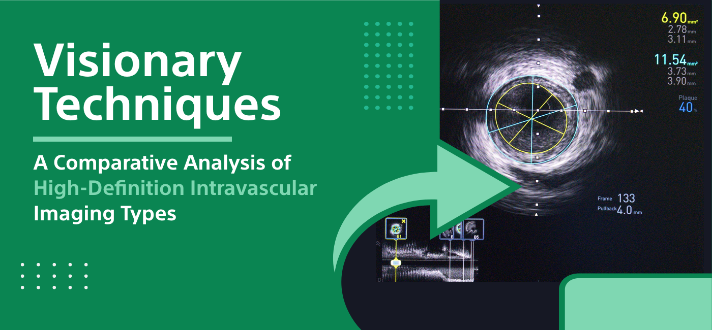
A Comparative Analysis of High-Definition Intravascular Imaging Types

Strong 8k brings an ultra-HD IPTV experience to your living room and your pocket.
Intravascular imaging is an ultrasound method of examining the interior of the blood vessels and the heart by using a tiny optical coherence tomography (OCT) probe or a special ultrasound probe inserted into the body. Eg: This method is the leading tool for guiding the interventional cardiologists and vascular surgeons during catheter-based procedures.
As a result of the technological achievements in this realm, some of the most sophisticated high-resolution intravascular imaging modalities have been brought forth, which serve for the more precise assessment of atherosclerotic coronary arteries and enhanced development of percutaneous coronary interventions (PCI).
I will cover the Types of High-Definition Intravascular Imaging.
In clinical practice, there are two type of high-definition intravascular imaging devices - Optical Coherence Tomography (OCT) and High-Definition Intravascular Ultrasound (B-II-VUS).
Optical Coherence Tomography
Using near-infrared light waves with high resolution, OCT produces cross sectional images of the vessel wall and plaques. It provides 10-fold sharper diagnostic accuracy than routine intravascular ultrasound.
The axial resolution of OCT is about 10-15 micrometers that enable high precision observation of the internal structure of atherosclerotic plaques including measurement of plaque thickness, fibrous cap thickness, and detection of features like cholesterol crystals, erosion, rupture, etc. It also efficiently measures stent apposition and coverage of the stent which culminates
In OCT, the flow of blood must be cleared from the lumen by flushing with saline in order to eliminate hemoglobin which blocks the light beams. The wires are coated with a catheter sheath which is Radio-Opaque. These markers help the radiologist identify artifacts in x-ray images. We pre-position the probe at the region of interest and then utilize automated pullback which enables us to produce longitudinal images.
High-Definition Intravascular Ultrasound
HD-IVUS utilizes a high-frequency (40-60 MHz) ultrasound waves to produce high-resolution cross-sectional 3D-images allowing to visualize anatomical features that would not be visible to the standard IVUS.
The technology is based on sophisticated approaches such as the combined backscatter technique, Virtual Histology and iMAP that make it possible for phased array to characterize tissue effectively and conduct a detailed plaque analysis.
HD-IVUS can provide the exact measurement of plaque burden and evaluate the features like calcification, necrosis etc. It can detect stenosis formation by thin-cap fibroatheroma, the most vulnerable plaques likely to rupture. Clear internal-external boundary cells are present for lumen and external elastic membrane.
This completely submerged phased-array mobile IVUS is capable of real-time focus shift and rendering of a series of three-dimensional images for lesion characterization.
Clinical Applications
Assessment of Coronary Atherosclerosis
Both OCT and HD-IVUS offer great resolution, which enables observation of coronary atherosclerotic lesions. In the way that this gives the exact determination for the plaque features such as thickness, composition, presence of thrombosis, erosion or rupture, these features serve an important role for the acute coronary events.
They can be most effectively used for determining whether a vulnerable plaque is present and assessing the amount of plaque, especially during acute coronary syndromes which require immediate treatment.
Facilitation of Percutaneous Coronary Interventions by the Guidance
The imaging technologies are utilized to sense stent sizes and positions which are later acted upon during the PCI procedures. Intravascular imaging has a significant role in decision-making for a substantial number of cases where PCI is concerned.
Post-intervention imaging, which can be used to measure reference vessel diameters along with appropriate stent sizing and landing zone selection, is done before the procedure. Another important component is the vulnerability plaque which decide the degree of heterogeneity and calcification; these are factors that determine tools to use.
After the stent implantation, the images will be examined to see the stent expansion, apposition and edge dissection. Targeting accuracy and the inclusion or exclusion of the lesion are the other aspects that are being considered to enable additional actions if necessary. It becomes the case that it leads to better long-term results in the end.
Follow-up and Surveillance
After surpassing a stent in the coronary artery, patients are often asked to undergo medical management and lifestyle modifications. Nevertheless, a few patients could get into In-Stent restenosis that would need repeat revascularization, which takes place with the repeat.
Through intravascular imaging, the stented segment and persistent segments are able to be documented, using systematic observation. This helps in objective qualification of the restenosis at the time of the follow-up angiograms.
Moreover it allows the visualization of the theory of restenosis – in-stent restenosis, stent fracture or incomplete stent expansion. This demand for such knowledge will help to choose the applicable therapy of balloon angioplasty, drug-eluting balloon or stent will be selected etc.
Evaluation of Bioresorbable Stents
Bioresorbable intravascular stent (BVS) were developed as the last innovation in totaling obstructive coronary artery depression. During this 2-3 years period, the scaffold disappears leaving behind the reversed vascular normal function with no other foreign bodies.
Intravascular imaging is a vital tool used for patient selection and individualization of BVS deployment. While doing the follow-up, it is tracking this process to help the doctor take the next steps of the procedure.
Fusion Imaging
Collocation of OCT and IVUS scans along with their fusing into a dataset with information about anatomy and composition resulted in a better diagnostic value that exceeds the quality of either method alone.
Thorough plaque characterization finding by the hybrid imaging of OCT/IVUS can not only achieve the improvement of stent optimization but also better the understanding of the stent failure mechanism.
Machine Learning
In addition, the artificial intelligence (AI) and convolutional neural networks have made a conscientious analysis of intracoronary imaging runs that include identifying key frames and quantifying of parameters such as lumen area or stent dimensions.
Machine learning algorithms have a high probability of changing this way of processing and interpreting these huge amounts of data to let us faster and more reliable analyses.
Molecular Imaging
A future mark of progress in the intravascular imaging will come with the development of specific fluorescent markers that will aid in visualizing inflammation level, protease activity, thrombosis, and neoangiogenesis which will enhance capability of this technology in finding vulnerable plaques or patients.
Read also: Understanding Atrial Fibrillation: Key Facts for Heart Health
Transluminal imaging is therefore not limited to anatomy only but it has great potential to transform this information into pathophysiology and even molecular level assessment of atherosclerosis in coming years
Conclusion
Diagnostic imaging of coronary blood vessels is achieved using high-definition technology including OCT and IVUS, which is known to be the gold standard for identifying cardiovascular pathology. With this technology there is additional a complimentary technique which has already confirmed clinical utility in the various types of patients from primary interventions to stent failures.
The continuous advancement and incorporation of intra-vascular imaging with innovative indices of plaque vulnerability can greatly modify the management of plaques of high-risk and patients both. It is possible for the patient outcomes to be improved by the development of the broad-based use of these methods.
Note: IndiBlogHub features both user-submitted and editorial content. We do not verify third-party contributions. Read our Disclaimer and Privacy Policyfor details.


