Biopsy for Diagnosis: Clear Facts, Steps, Risks, and Timelines
-

- March 15th, 2026
- 664 views

FREE SEO Topical Map Generator: Find Your Next Content Ideas
Biopsy for diagnosis: an essential guide
A biopsy for diagnosis is a medical procedure that removes tissue or cells to determine whether disease is present. This guide explains how biopsies work, common types, what to expect before and after the procedure, how results are reported, and practical steps to prepare. It is written for people seeking clear, actionable facts about biopsy testing and its role in diagnosis.
- Biopsy takes a small tissue sample for microscopic analysis (histology or cytology).
- Common methods: fine-needle aspiration, core needle biopsy, excisional biopsy, and image-guided biopsies.
- Risks are usually low but include bleeding, infection, and inconclusive results.
- Expect results in days to weeks depending on tests; immunohistochemistry and molecular tests add time.
Detected intent: Informational
What is a biopsy and why is it done?
A biopsy is the removal of tissue or cells from the body for microscopic examination by a pathologist. The main goals are to confirm or rule out cancer, identify infection or inflammation, classify disease subtypes, and guide treatment decisions. Biopsy findings provide definitive evidence that imaging or blood tests alone often cannot deliver.
Related terms and how the test is read
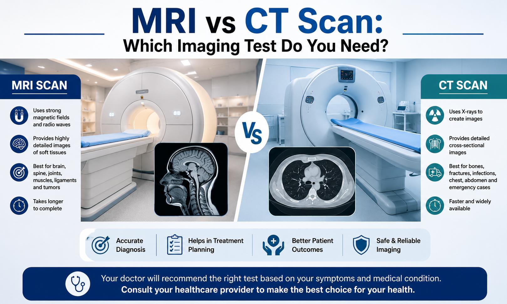
Pathology reports may use terms like histology, cytology, grade, stage, margins, and immunohistochemistry. Imaging guidance (ultrasound, CT, MRI) is often used to target areas precisely. For many conditions, a biopsy result is considered the diagnostic gold standard by clinical practice guidelines and specialty societies.
Types of biopsy procedures
Fine-needle aspiration (FNA)
FNA uses a thin needle to withdraw cells or fluid. It is quick and minimally invasive but may not capture tissue architecture needed for some diagnoses.
Core needle biopsy
Core biopsy removes a small cylinder of tissue with a larger hollow needle. It preserves tissue structure, improving diagnostic accuracy in solid organs and breast lesions.
Excisional and incisional biopsies
An excisional biopsy removes the entire suspicious area (common for skin or small lumps); an incisional biopsy removes a portion for diagnosis when full removal is not feasible at first.
Image-guided and surgical biopsies
Interventional radiologists often perform biopsies under ultrasound, CT, or MRI guidance for deep organs. Surgical biopsies occur in the operating room when tissue access is complex.
How results are reported and the biopsy results timeline
Typical pathology results are reported within a few days to a week. Standard histology is faster; specialized tests such as immunohistochemistry, genetic panels, or molecular profiling extend the biopsy results timeline to 1–3 weeks or longer. If an initial sample is inadequate, repeat biopsy may be necessary.
Preparing for a biopsy and what to expect
Pre-procedure instructions usually cover medication adjustments (anticoagulants), fasting for some procedures, and consent. Expect local anesthesia for many biopsies; sedation or general anesthesia is used for surgical biopsies. Post-procedure instructions include wound care, activity limits, and signs of complications to watch for.
Biopsy recovery time
Recovery varies by method: FNA and many core biopsies allow same-day discharge and minimal downtime (24–48 hours). Surgical excision requires longer recovery, often several days to weeks depending on the site and extent of surgery. Follow provider instructions for wound care and activity restrictions.
CLEAR Biopsy Checklist (framework)
Use the CLEAR checklist to prepare and evaluate any planned biopsy:
- Consent: Confirm informed consent and understanding of purpose and risks.
- Location: Verify the target site and imaging guidance plan.
- Extraction method: Confirm needle type, core vs aspiration, or surgical approach.
- Analysis plan: Specify required tests (histology, immunohistochemistry, molecular assays).
- Risks explained: Review bleeding, infection, and follow-up steps.
Practical tips before and after a biopsy
- Bring a list of medications and confirm whether blood thinners should be paused with the clinician.
- Arrange transportation if sedation is planned; avoid driving for 24 hours after anesthesia.
- Follow wound-care instructions precisely and call the clinic for fever, uncontrolled bleeding, or severe pain.
Practical tips: 3–5 actionable points
- Ask the clinician which exact tests the pathology lab will run so additional sampling can be requested at the time of biopsy.
- Confirm whether imaging guidance is necessary to improve yield and reduce the need for repeat procedures.
- Keep a copy of the pathology report and request clear follow-up steps from the care team.
Common mistakes and trade-offs
Choosing a biopsy method involves trade-offs between invasiveness, diagnostic yield, and risk. Common mistakes include:
- Undersampling—using FNA when a core biopsy is needed to assess tissue architecture.
- Not planning for ancillary tests—failing to request extra tissue for molecular or genomic testing can delay care.
- Ignoring anticoagulation guidance—continuing blood thinners increases bleeding risk.
Real-world example
Scenario: A patient presents with a 1.5 cm suspicious breast lump on mammogram. The team schedules an ultrasound-guided core needle biopsy. Local anesthesia and ultrasound guidance ensure accurate sampling; the specimen is sent for histology and receptor testing. Results arrive in 5 days confirming invasive carcinoma and hormone receptor status, guiding definitive surgical and medical management.
Safety, standards, and where to learn more
Clinical guidelines from pathology and specialty societies set best practices for biopsy technique and specimen handling. For clear patient-facing information on when and how biopsies are used, see this resource from the American Cancer Society for further background and references: American Cancer Society.
Core cluster questions
- How is a core needle biopsy different from fine-needle aspiration?
- When is an image-guided biopsy recommended?
- How much tissue is needed for molecular testing from a biopsy?
- What are the signs of infection or complications after a biopsy?
- How should anticoagulant medications be managed before a biopsy?
Next steps after receiving biopsy results
Review the pathology report with the ordering clinician. If diagnosis is confirmed, confirm staging tests and treatment options. If the sample is inadequate or results are inconclusive, discuss repeat biopsy or alternative diagnostic methods with the care team.
When to ask for a second opinion
Second opinions are appropriate when pathology reports are uncommon, when treatment decisions are consequential, or when molecular testing results will direct high-risk therapies. Pathology review by a subspecialist can clarify rare or complex findings.
FAQ: How long does a biopsy for diagnosis take?
Timing varies: the procedure itself can take 15 minutes to a few hours depending on complexity and anesthesia. Pathology reports typically return in days to a week, but specialized tests may extend the biopsy results timeline to several weeks.
FAQ: Are biopsies painful?
Local anesthesia minimizes pain during most biopsies. Mild discomfort or soreness at the site is common afterward; stronger pain control is used for surgical biopsies.
FAQ: What are biopsy risks and how common are complications?
Risks include bleeding, infection, bruising, and, rarely, damage to nearby structures. Complication rates differ by site and method; clinicians will discuss specific risks and mitigation strategies before the procedure.
FAQ: Can a biopsy miss cancer?
Yes. Sampling error or small lesions can produce false negatives. If clinical suspicion remains high, repeat biopsy or alternative diagnostic methods may be recommended.
FAQ: How should results be shared and stored?
Request a copy of the pathology report for personal records and share it with treating specialists. Digital patient portals often provide secure access to results.