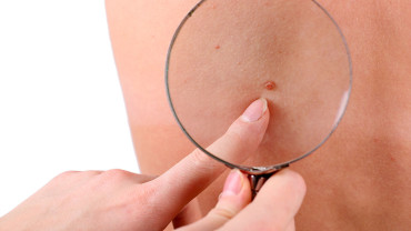
Dermoscopy in Dermatology: Revolutionizing Mole Evaluation Practices

Strong 8k brings an ultra-HD IPTV experience to your living room and your pocket.
Understanding Dermoscopy: A Comprehensive Guide to Mole Evaluation
Dermoscopy, a non-invasive skin imaging technique, has revolutionized the way medical professionals evaluate moles and other skin lesions. This comprehensive guide aims to demystify Dermoscopy Mole Evaluation in Dubai, making it accessible and easy to understand for anyone interested in learning more about this vital tool in dermatology.
What Is Dermoscopy?
Dermoscopy, also known as dermatoscopy, is a diagnostic method that allows the examination of skin lesions with greater clarity than the naked eye can provide. It involves using a specialized instrument called a dermatoscope, which magnifies the skin and illuminates it with polarized or non-polarized light. This method enables doctors to see structures and patterns within the skin that are not visible under normal light conditions.
Why Is Dermoscopy Important?
Dermoscopy is crucial in the early detection of skin cancer, particularly melanoma, the deadliest form of skin cancer. By enhancing the visualization of skin lesions, dermoscopy allows dermatologists to distinguish between benign (non-cancerous) and malignant (cancerous) moles more accurately. Early detection is key to successful treatment, making dermoscopy an essential tool in dermatology.
The Science Behind Dermoscopy
Dermoscopy works by reducing the surface reflection of the skin, allowing the observer to view subsurface structures. The dermatoscope uses different types of light, including polarized and non-polarized light, to achieve this. Polarized light helps in visualizing deeper skin layers, while non-polarized light is useful for seeing surface structures.
The tool also comes with magnification options, usually ranging from 10x to 20x, which helps in analyzing the intricate details of moles and other skin lesions. These details include pigment networks, dots, streaks, and other patterns that can indicate the nature of the lesion.
Key Features Observed in Dermoscopy
When evaluating a mole using dermoscopy, dermatologists look for specific features that help them assess the lesion. These features include:
Pigment Network: This is the network of lines seen in a mole, which can indicate whether a mole is benign or malignant. An irregular or atypical pigment network may suggest melanoma.
Dots and Globules: These are small, round structures within the mole. Their size, shape, and distribution can provide clues about the mole's nature.
Streaks: Streaks are radial lines that can be seen at the edge of a mole. Irregular streaks can be a warning sign of melanoma.
Blue-White Veil: A blue-white area within a mole can indicate a deeper lesion, which is often associated with melanoma.
Vascular Patterns: The presence of blood vessels in a certain pattern can also help differentiate between benign and malignant lesions.
How Is Dermoscopy Performed?
Dermoscopy is a relatively simple and quick procedure. The dermatologist applies a liquid (usually oil, alcohol, or water) to the skin lesion, which reduces surface reflection. The dermatoscope is then placed directly on the skin to get a clear view of the lesion.
Some dermatoscopes are handheld, while others are attached to a camera for capturing and storing images for further analysis. This allows for tracking changes in the lesion over time, which is crucial for early detection of skin cancer.
The ABCDE Rule in Dermoscopy
A widely used method in dermoscopy is the ABCDE rule, which helps in the initial evaluation of moles:
A for Asymmetry: If one half of the mole does not match the other, it could be a warning sign.
B for Border: Irregular, scalloped, or poorly defined borders are concerning.
C for Color: Multiple colors or an uneven distribution of color can indicate malignancy.
D for Diameter: Moles larger than 6mm (about the size of a pencil eraser) are more likely to be melanoma.
E for Evolution: Any change in size, shape, or color of a mole is a red flag.
The Role of Dermoscopy in Early Detection
Early detection of melanoma dramatically increases the chances of successful treatment. Dermoscopy plays a critical role in this by providing a clearer and more detailed view of moles, allowing for the identification of potentially malignant lesions at an earlier stage than would be possible with the naked eye alone.
Limitations of Dermoscopy
While dermoscopy is a powerful tool, it is not infallible. The accuracy of dermoscopy depends heavily on the experience and expertise of the dermatologist. False positives and false negatives can occur, which is why dermoscopy should be used in conjunction with other diagnostic methods, such as biopsy, for a definitive diagnosis.
The Future of Dermoscopy
The future of dermoscopy looks promising, with advancements in digital dermoscopy and artificial intelligence (AI) enhancing its capabilities. Digital dermoscopy involves capturing high-resolution images of skin lesions and storing them for comparison over time. AI, on the other hand, is being developed to assist in the analysis of these images, potentially increasing the accuracy of mole evaluations.
Conclusion: The Importance of Regular Skin Checks
Dermoscopy is an invaluable tool in the early detection of skin cancer, particularly melanoma. By providing a detailed view of skin lesions, it allows for more accurate assessments, leading to early intervention and better outcomes. Regular skin checks, both self-examinations and professional evaluations, are crucial in maintaining skin health. Understanding dermoscopy and its role in mole evaluation empowers individuals to take proactive steps in monitoring their skin and seeking medical advice when needed. Early detection saves lives, and dermoscopy is at the forefront of this effort.
Note: IndiBlogHub features both user-submitted and editorial content. We do not verify third-party contributions. Read our Disclaimer and Privacy Policyfor details.


