How MRI Works: A Clear Guide to Magnetic Resonance Imaging and Safety
-
- March 04th, 2026
- 327 views
Magnetic resonance imaging (MRI) is a noninvasive medical imaging method that uses magnetic fields and radiofrequency energy to produce detailed pictures of internal anatomy. This article explains how MRI works, what makes the images detailed and safe, and practical steps patients and clinicians can take to reduce risk and improve image quality.
Detected intent: Informational
Key points: MRI uses a strong magnet and radiofrequency pulses to align and read signals from hydrogen protons. Safety relies on screening for metal, understanding MRI safety features, and using protocols optimized for the clinical question.
How MRI Works: Basic Physics and Image Formation
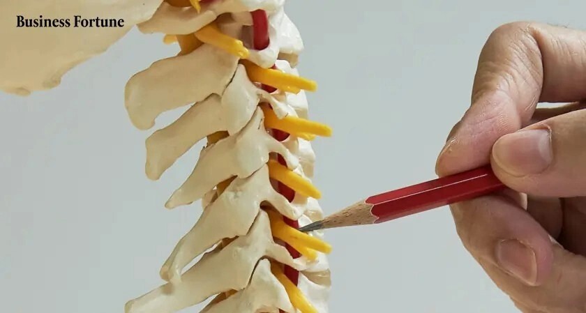
The core idea of how MRI works is resonance of hydrogen nuclei in the body. Human tissue contains abundant hydrogen atoms in water and fat. When a strong static magnetic field (B0) is applied, these hydrogen protons align with or against the field. Short bursts of radiofrequency (RF) energy tuned to the proton Larmor frequency briefly knock that alignment out of equilibrium. When the RF pulse stops, protons relax back to alignment and emit measurable signals. Gradient magnetic fields localize those signals so the scanner can reconstruct a 3D image using Fourier transform math.
Key technical terms
- T1 and T2 relaxation times — how quickly protons recover longitudinal or lose transverse magnetization; they determine tissue contrast.
- Gradient coils — create spatial variations in the magnetic field, enabling slice selection and image encoding.
- RF coils — transmit pulses and receive returning signals; coil design affects signal-to-noise ratio (SNR).
- Field strength — commonly 1.5T and 3T in clinical imaging; higher field often yields better SNR but introduces trade-offs.
MRI safety features and what keeps MRI safe
Modern scanners incorporate multiple MRI safety features to minimize risk. These include controlled access zones, patient monitoring systems, interlock mechanisms that halt RF and gradients if unsafe conditions arise, and protocols for contrast administration. Screening procedures identify implanted devices or loose metal that could move or heat during scanning. Regulatory bodies and professional societies publish safety guidance used by imaging departments.
For authoritative safety recommendations and device information, consult the U.S. Food and Drug Administration’s MRI safety resources: FDA: Magnetic Resonance Imaging (MRI).
MRI S.A.F.E. checklist (named framework for practical use)
The MRI S.A.F.E. checklist organizes the most important pre-scan and in-scan steps into a simple framework suitable for clinical teams and informed patients.
- Screen — Verify medical history, implants, and metal; use written screening forms and verbal confirmation.
- Assess — Check implant labeling (MR-safe, MR-conditional, MR-unsafe) and consider device-specific conditions.
- Field-strength selection — Choose 1.5T vs 3T or specialized low-field for implants, pediatric exams, or specific sequences.
- Explain — Communicate expected sensations (noise, warmth), contrast risks, and motion-reduction techniques to the patient.
- Follow-up — Document any adverse events, ensure post-contrast monitoring if used, and archive screening data.
Real-world example: diagnosing a torn meniscus
Scenario: A 35-year-old athlete presents with knee pain after a pivot injury. The magnetic resonance imaging process begins with screening for metal and recent surgeries. A dedicated knee coil is positioned to maximize SNR. Sequences chosen include proton-density with and without fat suppression to highlight meniscal tears and fluid-sensitive T2-weighted sequences to show edema. The tech instructs the patient to remain still and uses padding to reduce motion. Images are reviewed for meniscal morphology and associated ligament or cartilage injuries. If diagnostic clarity is insufficient, a targeted repeat sequence at higher resolution may be acquired, balancing additional scan time against patient comfort.
Practical tips for patients and clinicians
- Bring or disclose all implant cards and surgical reports; many implants are conditionally safe only under certain scanner settings.
- Remove all external metal: jewelry, clothing with metal fasteners, hearing aids, and credit cards before entering the scan room.
- Use ear protection; MRI machines produce high-decibel gradient noise during sequences.
- For improved image quality, limit motion by practicing breath holds when instructed and using cushions or straps recommended by staff.
- Discuss contrast risks with the ordering clinician if there is a history of kidney disease; contrast agents like gadolinium are common but require assessment.
Common mistakes and trade-offs when using MRI
Trade-offs are inherent in selecting MRI parameters. Higher field strength (3T) improves signal-to-noise ratio and spatial resolution but increases susceptibility artifacts, RF heating risk, and specific absorption rate (SAR). Longer scan protocols produce more detailed images but raise motion artifact risk and reduce throughput. Overlooking implant labeling or failing to screen thoroughly are common mistakes that can cause patient harm. Choosing the wrong coil or sequence can miss subtle pathology; match the protocol to the clinical question.
Core cluster questions
- What is the difference between T1 and T2 MRI images?
- How do MRI contrast agents work and when are they necessary?
- What safety screening is required before an MRI scan?
- How does field strength (1.5T vs 3T) affect image quality and safety?
- What steps reduce motion artifacts during an MRI scan?
Practical imaging considerations for clinicians
Align the imaging protocol with the clinical question: vascular windows for angiography, fat-suppressed sequences for marrow pathology, or diffusion-weighted imaging for acute stroke. Calibration and periodic quality assurance, as recommended by professional societies such as the American College of Radiology, ensure reliable performance. Document MR-conditional implant parameters and follow the device manufacturer’s conditions carefully.
FAQ
How MRI works: is the procedure safe for adults and children?
Yes, MRI is generally safe for adults and children when proper screening and safety protocols are followed. There is no ionizing radiation. Risks primarily relate to metal in the body, implanted devices, heating from RF energy, and contrast agent side effects. Pediatric imaging may require sedation in some cases; specialists weigh benefits and risks before proceeding.
Can implants like pacemakers or cochlear implants go into an MRI?
Some implants are labeled MR-safe, MR-conditional, or MR-unsafe. MR-conditional implants may be scanned under specific conditions (field strength, scanner model, positioning). Always verify implant details with device documentation and consult radiology or cardiology teams if needed.
Why do certain scans use contrast agents?
Gadolinium-based contrast agents change local magnetic properties and improve visualization of blood vessels, inflammation, tumors, and regions with abnormal blood-brain barrier. Contrast is used when it provides diagnostic value beyond non-contrast sequences and when renal function permits safe administration.
How long does an MRI scan typically take?
Scan times vary from 10–15 minutes for a focused study to 45–60 minutes for comprehensive exams. Longer protocols that require higher resolution or multiple planes increase diagnostic detail but also risk patient motion; protocol optimization balances time, quality, and clinical need.
What should a patient do if they feel claustrophobic in the scanner?
Inform staff before the scan. Options include open-bore scanners, shorter breath-hold sequences, audio/visual systems, mild anxiolytics prescribed by a physician, or in some cases sedation. Communication during the exam and training with mock scanners can help reduce anxiety.
Related entities and synonyms used: magnetic resonance imaging, MRI physics, hydrogen protons, gradient coils, RF coils, T1/T2 relaxation, gadolinium contrast, specific absorption rate (SAR), field strength, image reconstruction, Fourier transform, signal-to-noise ratio (SNR).