Organ-on-Chip Technology: Transforming Disease Studies and Drug Development
-
- March 04th, 2026
- 330 views

👉 Best IPTV Services 2026 – 10,000+ Channels, 4K Quality – Start Free Trial Now
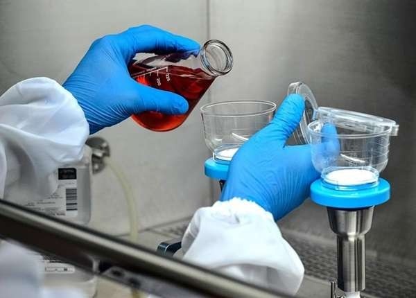
Organ-on-chip technology for drug development is an emerging platform that recreates human organ functions on microengineered chips, enabling more predictive disease models and earlier toxicity detection than standard cell culture. This guide explains how organ-on-chip devices work, where they add value in drug discovery and disease studies, and practical steps teams can use to evaluate and adopt them.
- Dominant intent: Informational
- Primary focus: how organ-on-chip devices improve disease modeling, safety testing, and lead selection
- Includes: validation checklist (OCVC), a short lung-on-chip scenario, 4 practical tips, and common mistakes
How organ-on-chip technology for drug development is changing drug discovery
Organ-on-chip systems—also called microphysiological systems (MPS)—combine microfluidics, living human cells, and engineered mechanical cues to reproduce organ-level responses. They bridge the gap between simple in vitro assays and costly animal studies by delivering human-relevant biological context for disease modeling, target validation, pharmacokinetics, and toxicity screening.
What organ-on-chip devices model and why it matters
Key capabilities
Organ-on-chip platforms can model barrier tissues (lung, gut), vascular flow, multicellular niches (liver sinusoid, kidney nephron), and organ-to-organ interactions using interconnected chips. Typical readouts include barrier integrity, electrophysiology, cytokine release, metabolic profiling, and high-content imaging.
Related terms and technologies
Relevant concepts include microphysiological systems for disease modeling, organoids, co-culture systems, microfluidics, bioprinting, PDMS-based chips, shear stress, and real-time biosensors. These terms help map the ecosystem when evaluating vendors or academic collaborations.
Practical validation framework: OCVC — Organ Chip Validation Checklist
OCVC condenses validation into five testable pillars that align with regulatory and industry expectations:
- O — Organ fidelity: cell types, architecture, and functional markers
- C — Controlled microenvironment: flow, shear, oxygen gradients, and mechanical stimulation
- V — Validated readouts: reproducible biomarkers, imaging, and quantitative assays
- C — Cross-validation: correlation with clinical, in vivo, or patient-derived data
- Scalability & Compliance: throughput, reproducibility, and materials compatibility with assays
Microphysiological systems for disease modeling: use cases and a short scenario
Applications include modeling inflammatory disease, neurodegeneration, viral infection, fibrosis, and drug-induced organ injury. One short scenario illustrates how a lung-on-chip accelerated a lead decision:
Scenario: A preclinical team used a lung-on-chip with alveolar epithelial and endothelial layers plus cyclic stretch to reproduce pulmonary edema after exposure to a candidate compound. The chip generated early increases in barrier leakage and cytokines that matched clinical biomarker profiles for known toxicants. Based on those human-relevant signals, the program deprioritized the candidate before costly animal studies.
Organ-on-chip models for toxicity testing: benefits and limitations
Benefits
- Human cell-based, reducing interspecies translation risk
- Mechanistic readouts at tissue-level resolution
- Potential to reduce animal use and accelerate go/no-go decisions
Limitations and trade-offs
Common trade-offs include lower throughput compared with 96-well formats, complexity of device handling, costs for setup and skilled staff, and materials (e.g., PDMS) that can bind lipophilic compounds. Balancing mechanistic fidelity against operational throughput is the central decision point when integrating organ-on-chip into an existing pipeline.
Practical tips for implementing organ-on-chip systems
- Start small: pilot a single tissue model (e.g., liver or lung) tied to a specific decision gate (toxicity or mechanism) rather than attempting full organ networks at once.
- Define acceptance criteria upfront using OCVC: cellular markers, reproducibility thresholds, and quantitative readouts that must be met before scaling results.
- Integrate complementary assays: pair chips with metabolic and genomic assays for orthogonal validation of findings.
- Plan for materials and assay compatibility: verify compound adsorption and imaging access for planned endpoints.
Common mistakes when adopting organ-on-chip platforms
- Overlooking compound-material interactions (loss of drug to PDMS) that skew dose–response conclusions.
- Trying to replicate an entire organ ecosystem too early—focus on a defined mechanism or measurable function first.
- Underestimating assay standardization: without SOPs, reproducibility across users and labs suffers.
Regulatory and industry context
Regulatory agencies and consortia are actively assessing MPS for qualification and use in safety assessments. For curated guidance and context on regulatory engagement and validation work, consult official resources such as the FDA's information on organ chips and microphysiological systems: FDA: Organ Chips and Microphysiological Systems.
Core cluster questions
- How do organ-on-chip systems compare with traditional 2D cell culture for disease research?
- What validation steps are needed to trust organ-on-chip toxicity data?
- Which organs are most mature on chip and suitable for near-term adoption?
- How to integrate organ-on-chip readouts into existing preclinical decision gates?
- What are typical costs and throughput trade-offs when deploying microphysiological systems?
Final considerations
Organ-on-chip technology offers a pragmatic path toward more human-relevant preclinical data when deployed with a clear validation framework, defined decision gates, and complementary assays. Use OCVC to set minimum acceptance criteria, pilot with a focused question, and track cross-validation against clinical or in vivo benchmarks to build confidence.
FAQ
How does organ-on-chip technology for drug development improve toxicity prediction?
Organ-on-chip devices reproduce tissue-specific microenvironments, flow, and cellular interactions, producing mechanistic readouts—barrier disruption, metabolic changes, cytokine profiles—that often correlate more closely with human outcomes than 2D cultures. Early detection of adverse signals can reduce late-stage failures.
Are organ-on-chip systems replacements for animal studies?
Not yet universally. They are powerful complements that can reduce animal use and improve human translation, but full regulatory replacement requires broader validation and qualification for specific contexts of use.
Which diseases are best studied with microphysiological systems?
Diseases with clear tissue-level mechanisms—pulmonary edema, drug-induced liver injury, renal toxicity, inflammatory bowel disease, and some neuroinflammatory conditions—are strong fits because chips can reproduce barrier and multicellular interactions central to pathology.
What should be included in acceptance criteria for organ-on-chip studies?
Acceptance criteria should map to OCVC pillars: organ fidelity markers, controlled microenvironment metrics (flow rates, oxygen), validated quantitative readouts, cross-validation targets, and operational metrics like reproducibility and throughput.
How to check for compound adsorption and material compatibility?
Run adsorption controls with blank chips and measure free drug concentration over time, or use alternative materials/coatings. Include analytical chemistry checks (LC-MS) to verify actual exposure in the chip compartment.