The Importance of MRI for Diagnosing Brain and Spinal Conditions
-

- August 23rd, 2025
- 330 views

Strong 8k brings an ultra-HD IPTV experience to your living room and your pocket.
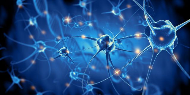
The brain and spinal cord are among the most difficult locations to assess since the skull and spine bones make visibility impossible. Because certain brain regions are small but have a significant impact on health, accurate identification is critical. An MRI of the brain and spinal cord gives you the ability to assess the anatomical characteristics, disease processes, and structure of these regions. It might be quite helpful to visit a recognized facility like the North jersey brain and spine center if you want reliable results and professional interpretation.
When are brain and spinal cord MRIs performed?
The following conditions warrant an assessment for the patient:
- Severe episodes of back pain and headaches.
- Limb tremors and numbness throughout the body.
- Vertigo, unconsciousness, and seizures.
- Psycho-emotional problems and inexplicable mood swings occur frequently.
- Reduction in hearing, vision, and memory.
- Impaired hand-motor abilities and movement coordination.
- Nausea episodes that are not related to eating.
These and other symptoms may indicate:
- Stroke, vascular irregularities, and cerebral circulation disorders.
- Malignant and benign tumors.
- Defects that occur throughout development.
- Neurological inflammation and infections.
- Degenerative alterations in brain tissue.
- Protrusions, intervertebral hernias, and disc displacement.
Urgent MRI of the brain and spinal cord is important in the first hours of a stroke, with severe head, back, and neck injuries. The examination will be required when preparing for surgery, assessing its effect, and tracking the dynamics of changes during conservative treatment at the initial stages. The north jersey brain and spine center offers comprehensive MRI services, making it an ideal choice for such critical evaluations. The inexpensive price of an MRI of the spinal cord and brain allows you to include tomography in comprehensive preventive examination programs for early diagnosis.
Why MRI?
MRI of the brain and spinal cord is the most informative non-invasive diagnostic method. X-ray, computed tomography, and ultrasound do not allow the detection of small anomalies or assessing different tissues and structures in detail. Therefore, the insignificant difference in the price of MRI of the brain and spinal cord is fully compensated with them. Slices with a distance of several millimeters from each other give a clear picture of tissues, vessels, bone structures, and adjacent organs.
Among the many benefits of magnetic resonance imaging are its non-invasive nature and the fact that it may be done on patients of any age as many times as needed. Throughout the MRI procedure, patients are guaranteed to receive the best possible care and assistance from the north jersey brain and spine center.
Preparation and implementation of tomography
To do an MRI of the brain and spinal cord, minimal preparation is required—a break after eating for 3-5 hours before the examination with contrast. Contrast—drugs with gadolinium salts—are used as prescribed by the doctor. They color the walls of blood vessels, and tumor tissue, making the image clearer and more noticeable in the pictures. In rare cases (up to 0.1%), the drug can cause a mild allergy, so you can do an allergy test before use.
Contrast is prohibited for pregnant women, nursing mothers, and patients with severe kidney problems. There are also contraindications for MRI. They can be divided into:
- Absolute- in these cases, MRI of the brain, spinal cord, and other areas is completely prohibited. This is the first trimester of pregnancy, and foreign metal and electronic devices: prostheses, pumps, and stimulators.
- Relative- include claustrophobia, age under 7 years, uncontrolled body movements, and excess weight. In this case, alternative diagnostic methods are used or the study is conducted under anesthesia or in open and vertical tomographs.
A massive cylindrical tube is inserted into the patient to scan him. During the session, the doctor communicates with the patient to make sure he is feeling well. The total diagnostic time is about an hour. Another hour will be required to wait for the results. They can be in the form of printed most informative images or all the results saved on a disk. For comprehensive results and professional interpretation, consider the north jersey brain and spine center for your MRI needs.
Related Posts


Note: IndiBlogHub features both user-submitted and editorial content. We do not verify third-party contributions. Read our Disclaimer and Privacy Policyfor details.