Practical Risk Assessment with Dermoscopy for Effective Skin Cancer Screening
-

- March 20th, 2026
- 527 views

Get a free topical map and start building content authority today.
Dermoscopy in skin cancer screening improves lesion evaluation by revealing sub-surface structures not visible to the naked eye. This guide summarizes how to use dermoscopy for risk assessment, presents a concise checklist for triage, and explains common dermoscopic features linked to malignancy.
- Dermoscopy increases diagnostic accuracy for melanoma and non-melanoma skin cancers when combined with clinical context and follow-up.
- Use a structured checklist to classify risk and decide between immediate biopsy, short-interval follow-up, or routine monitoring.
- Be aware of limitations: operator experience, lesion variability, and false negatives for some amelanotic or nodular tumors.
Detected intent: Informational
Dermoscopy in skin cancer screening: risk assessment essentials
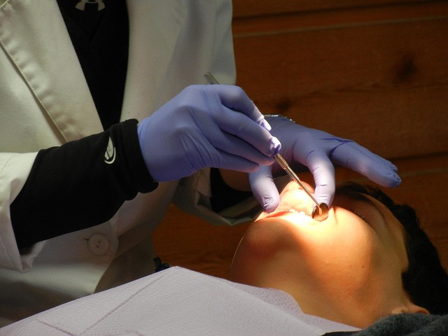
Dermoscopy is a noninvasive imaging technique that aids risk stratification during skin cancer screening. When used properly, dermoscopy improves sensitivity and specificity compared with naked-eye examination by revealing pigment networks, vascular patterns, and other diagnostic structures (polarized and non-polarized modes each highlight different features). National specialty bodies such as the American Academy of Dermatology endorse dermoscopic evaluation as part of standard assessment pathways for suspicious lesions.
Dermoscopy Risk Assessment Checklist (DRAC)
A short, repeatable framework helps convert visual dermoscopic findings into risk categories. The Dermoscopy Risk Assessment Checklist (DRAC) below is a practical model for clinics and primary care.
- Demographics & History: age, personal/family melanoma history, previous atypical nevi, recent change, bleeding, pain.
- Clinical Context: lesion location, sun-exposure pattern, patient skin type, immunosuppression status.
- Basic Dermoscopic Screen: symmetric vs asymmetric global pattern; presence of pigment network, globules, streaks, blue-white veil, regression structures.
- Red Flags: atypical vascular pattern, polymorphous vessels, structureless blue or white areas, irregular streaks, peripheral radial streaming.
- Risk Category: High (biopsy/excision), Intermediate (short-interval follow-up 6–12 weeks or diagnostic excision depending on clinical concern), Low (routine monitoring).
This checklist complements established diagnostic algorithms such as the ABCDE rule and the Seven-Point Checklist; DRAC focuses on triage decisions rather than definitive diagnosis.
How to interpret common dermoscopic features
Dermoscopic criteria for melanoma: key signs to look for
Important dermoscopic features associated with melanoma include asymmetric pigment networks, atypical pigment dots and globules, irregular streaks, blue-white veil, and regression structures (white scar-like areas and blue-gray granularity). Patterns differ by subtype (e.g., lentigo maligna, superficial spreading, nodular melanoma). Correlate dermoscopic features with clinical history and lesion evolution before assigning high-risk status.
Other entities and mimics
Non-melanoma skin cancers show different but sometimes overlapping dermoscopic findings: basal cell carcinoma commonly presents arborizing vessels and leaf-like areas; squamous cell carcinoma precursors may show keratin and scale with irregular vessels. Amelanotic lesions lack pigment clues and rely on vascular morphology and clinical suspicion.
Real-world scenario
A 58-year-old patient with a history of outdoor work presents with a 6-mm asymmetric brown macule on the forearm that reportedly darkened over 3 months. Dermoscopic evaluation shows an irregular pigment network, multiple irregular globules, and a small blue-white area. Applying DRAC, the lesion meets high-risk criteria: immediate diagnostic excision with histopathology is advised rather than watchful waiting.
Practical tips for accurate dermoscopy risk assessment
- Standardize imaging: take polarized and non-polarized dermoscopic images and label with orientation and date to support follow-up comparisons.
- Use serial monitoring for equivocal lesions: compare size, structure, and new dermoscopic features at defined intervals (6–12 weeks for suspicious change).
- Combine clinical and dermoscopic data: use history of change, patient risk factors, and dermoscopic findings together—never rely on dermoscopy alone.
- Document decisions and communicate risk clearly with patients, using visual aids when possible to explain rationale for biopsy vs surveillance.
Trade-offs and common mistakes
Common mistakes
- Over-reliance on single dermoscopic signs without clinical context, leading to unnecessary biopsies.
- Underestimating amelanotic or nodular lesions because they lack classic pigment patterns—vascular assessment is critical.
- Poor image quality or inconsistent follow-up intervals that obscure subtle evolution.
Trade-offs include sensitivity versus specificity: lowering the threshold for biopsy reduces missed cancers but increases benign excisions. The decision should balance patient risk factors, cosmetic considerations, and access to specialist pathology.
Core cluster questions
- How does dermoscopy change the diagnostic accuracy for melanoma compared with naked-eye examination?
- What are the most predictive dermoscopic features for early melanoma?
- When is short-interval dermoscopic follow-up appropriate versus immediate excision?
- How should amelanotic lesions be assessed with dermoscopy?
- What training and quality controls improve dermoscopy performance in primary care?
Practical implementation checklist
Use this short checklist before concluding risk:
- Confirm patient risk factors and lesion history.
- Acquire high-quality polarized and non-polarized images.
- Apply DRAC: screen, flag red signs, assign risk category.
- Decide: biopsy, short-interval monitoring, or routine follow-up; document plan.
- Refer to dermatology for uncertain high-risk lesions or diagnostic dilemmas.
FAQ
How sensitive is dermoscopy in skin cancer screening?
Sensitivity varies with operator experience and lesion type; meta-analyses show improved sensitivity and specificity for melanoma compared with naked-eye exam when dermoscopy is used by trained clinicians. Performance increases with structured algorithms and regular practice.
What are the limits of dermoscopy risk assessment?
Dermoscopy cannot replace histopathology. Limitations include operator dependency, overlap of features between benign and malignant lesions, and difficulty detecting certain amelanotic or nodular tumors.
When should suspicious findings on dermoscopy prompt biopsy?
Biopsy is indicated when dermoscopic evaluation reveals multiple high-risk features (irregular network, streaks, blue-white veil, atypical vessels) or when a lesion shows definite clinical change in a patient with risk factors. Use DRAC to standardize the threshold for biopsy.
How should dermoscopy in skin cancer screening be integrated into primary care?
Integrate dermoscopy by providing targeted training, standardized imaging protocols, a clear referral pathway to dermatology, and use of checklists like DRAC to reduce variability in decision-making.
What are reliable resources for learning dermoscopic criteria for melanoma?
Trusted resources include specialty society guidance and published diagnostic algorithms. Regular hands-on training and image-based case review improve accuracy over time.