Dermoscopy for Mole Diagnosis: A Practical Guide to Distinguishing Common and Aberrant Moles
-
- March 15th, 2026
- 510 views

👉 Best IPTV Services 2026 – 10,000+ Channels, 4K Quality – Start Free Trial Now
Introduction
Dermoscopy for mole diagnosis is a noninvasive, high-yield technique that improves the ability to distinguish common benign nevi from atypical or malignant lesions. This guide explains how dermoscopy fits into differential diagnosis, presents a practical checklist, shows a short clinical scenario, and lists concrete tips and common pitfalls for clinicians and informed patients.
- Detected intent: Informational
- Primary keyword: dermoscopy for mole diagnosis
- Secondary keywords: differential diagnosis of moles; ABCDE checklist for melanoma
- Named checklist: ABCDE checklist for melanoma
- Authoritative resource: American Academy of Dermatology guidance on melanoma
- Core cluster questions:
- How to use dermoscopy to differentiate a common nevus from an atypical nevus?
- Which dermoscopic patterns suggest melanoma vs benign lesions?
- When should a suspicious mole be biopsied instead of monitored?
- How does polarized light dermoscopy compare to non-polarized dermoscopy?
- What are the limits of dermoscopy and when is dermatopathology required?
Dermoscopy for mole diagnosis: when and how to use it
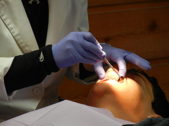
Dermoscopy augments naked-eye inspection by revealing subsurface structures — pigment networks, vascular patterns, and specific colors — that change the pre-test probability for melanoma and other diagnoses. Use dermoscopy when a lesion raises clinical concern (any evolving, symptomatic, or irregular mole) or during routine skin exams for patients with many nevi or a history of skin cancer.
Key concepts and terminology
Common and aberrant moles
Common nevi (junctional, compound, intradermal) typically show symmetric patterns and uniform color. Aberrant moles include dysplastic nevi and Spitz nevi, which may display asymmetry, atypical pigment networks, or irregular vascular features. Melanoma often breaks established patterns, showing chaotic structure, multiple colors, and atypical vessels.
Dermoscopy patterns and terms
Recognized terms include pigment network, globules, streaks, regression structures, blue-white veil, and atypical vascular patterns. Pattern analysis and algorithmic approaches (e.g., the 7-point checklist, the 3-point checklist) assist consistency in interpretation.
Practical diagnostic framework: ABCDE checklist + pattern analysis
Combine the named ABCDE checklist (Asymmetry, Border irregularity, Color variegation, Diameter >6 mm, Evolving) with dermoscopic pattern analysis to prioritize lesions for biopsy. The ABCDE checklist functions as a clinical triage tool; dermoscopy refines the differential diagnosis.
ABCDE checklist mapped to dermoscopic signs
- A — Asymmetry in both structure and color under dermoscopy
- B — Irregular, notched, or poorly defined borders with peripheral streaks
- C — Multiple colors (brown, black, blue-gray, red/white areas)
- D — Rapid change or large size; consider dermoscopy for internal heterogeneity
- E — Evolution in size, shape, color, or symptoms (itch, bleed)
Short clinical scenario
A 42-year-old patient presents with a new pigmented lesion on the forearm that has darkened over 3 months. Clinical exam shows slight asymmetry. Dermoscopy reveals an atypical pigment network with irregular streaks and a blue-gray regression area. Using the ABCDE checklist and dermoscopic pattern analysis, the lesion meets criteria for excisional biopsy and histopathologic evaluation. This pathway avoids unnecessary delay while minimizing unnecessary biopsies of clearly benign lesions.
Practical tips for imaging and interpretation
- Use both polarized and non-polarized dermoscopy when possible: polarized mode enhances deeper structures, non-polarized highlights surface features.
- Document baseline dermoscopic images for all at-risk patients to support serial comparisons and detect evolution.
- Apply an algorithm (for example, the 3-point checklist for quick triage) when time is limited; escalate to pattern analysis for indeterminate cases.
- When in doubt, prefer excisional biopsy with narrow margins rather than prolonged monitoring for lesions that score positively on ABCDE or show chaotic dermoscopic features.
Common mistakes and trade-offs
Common mistakes
- Over-reliance on dermoscopy alone: dermoscopy improves sensitivity but must be integrated with history and clinical exam.
- Poor image documentation: without baseline images, subtle evolution is missed.
- Misapplication of algorithms: checklists are aids, not absolute rules; atypical presentations occur.
Trade-offs
Using dermoscopy reduces unnecessary biopsies but can increase short-term surveillance workload. Broad use of low-threshold biopsy increases diagnostic yield at the cost of more benign excisions. Balance depends on patient risk factors, cosmetic concerns, and clinical setting.
Limits and escalation: when dermatopathology is required
Dermoscopy cannot provide a definitive histologic subtype. Any lesion with persistent atypical dermoscopic features, symptomatic change, or clinical concern should be biopsied. Consult dermatopathology for ambiguous histology; correlate dermoscopic images with pathology to reduce discordance.
Core cluster questions for further reading and internal linking
- How to use dermoscopy to differentiate a common nevus from an atypical nevus?
- Which dermoscopic patterns suggest melanoma vs benign lesions?
- When should a suspicious mole be biopsied instead of monitored?
- How does polarized light dermoscopy compare to non-polarized dermoscopy?
- What are the limits of dermoscopy and when is dermatopathology required?
Practical implementation checklist
Use this short checklist to operationalize dermoscopy in practice:
- Obtain high-quality baseline dermoscopic images for at-risk patients.
- Apply the ABCDE checklist during clinical exam.
- Use a rapid dermoscopic triage algorithm (3-point or 7-point) for time-limited cases.
- Biopsy any lesion meeting clinical or dermoscopic concern thresholds.
- Document outcomes and correlate with histopathology to improve accuracy.
When to refer
Refer complex or recurrent atypical lesions, discordant clinical-pathologic cases, or suspected melanoma to dermatology or a multidisciplinary skin cancer clinic for definitive management.
FAQ
Can dermoscopy for mole diagnosis replace a biopsy?
Dermoscopy cannot reliably replace histopathology when a lesion is suspicious. Dermoscopy improves pre-biopsy assessment and can reduce unnecessary biopsies, but definitive diagnosis of malignancy requires tissue sampling and pathology in cases with concerning clinical or dermoscopic features.
How accurate is dermoscopy at distinguishing melanoma from benign moles?
When used by trained examiners, dermoscopy significantly increases sensitivity and specificity compared with naked-eye exam. Accuracy depends on clinician experience, lesion type, and use of systematic algorithms. Continuous training and correlation with histopathology improve diagnostic performance.
What is the ABCDE checklist for melanoma and how does it relate to dermoscopy?
The ABCDE checklist (Asymmetry, Border, Color, Diameter, Evolving) is a clinical triage tool. Dermoscopy provides additional structural detail that maps to these criteria and can reveal asymmetry, border irregularity, and color variegation not visible to the naked eye.
How often should moles be monitored with dermoscopy?
Surveillance intervals depend on risk: high-risk patients (personal or family history of melanoma, many atypical nevi) often require annual or more frequent imaging, while low-risk patients may be monitored less frequently. Persistent change or new symptoms warrant earlier re-evaluation.
When is dermatopathology necessary after dermoscopy?
Any lesion with suspicious dermoscopic features, rapid evolution, symptomatic change, or discordant clinical assessment should be excised and submitted for dermatopathology. Pathology remains the reference standard for diagnosis.