Macrophages in Cancer: Isolation Methods, Functional Roles, and Clinical Insights
-
- February 23rd, 2026
- 1,277 views

👉 Best IPTV Services 2026 – 10,000+ Channels, 4K Quality – Start Free Trial Now
The term macrophages in cancer refers to immune cells that populate tumors and surrounding tissue and influence disease progression, therapy response, and tissue remodeling. Studies of macrophages in cancer combine laboratory isolation, molecular phenotyping, and functional assays to link cell state with tumor biology.
- Macrophages are abundant and heterogeneous components of the tumor microenvironment, often called tumor-associated macrophages (TAMs).
- Isolation methods include density gradient centrifugation, mechanical/enzymatic tissue digestion, magnetic and flow cytometry sorting.
- Phenotyping uses surface markers, cytokine profiling, single-cell transcriptomics, and spatial methods.
- Functional assays evaluate phagocytosis, cytokine production, interactions with cancer and stromal cells, and migration.
- Understanding macrophage states supports basic research and informs therapeutic strategies under clinical investigation.
Macrophages in cancer: overview of roles and heterogeneity
Tumor-associated macrophages (TAMs)
Tumor-associated macrophages (TAMs) are a major immune population within most solid tumors. TAMs influence angiogenesis, matrix remodeling, immune suppression, and responses to chemotherapy, radiation, and immunotherapy. Their effects vary with origin (tissue-resident versus monocyte-derived), microenvironmental cues, and temporal dynamics during tumor evolution.
Polarization and phenotypic diversity
Describing macrophages as strictly M1 (pro-inflammatory) or M2 (anti-inflammatory) is an oversimplification. Modern studies emphasize a spectrum of activation states defined by transcriptional programs, metabolic profiles, and functional outputs. Single-cell RNA sequencing and multiplex imaging have revealed multiple TAM subpopulations with distinct gene signatures and spatial niches.
Isolation and characterization of macrophages
Sources of cells
Macrophages for cancer research are obtained from human tumor resections, peripheral blood mononuclear cells (PBMCs), mouse tumor models, or engineered organoid/co-culture systems. Fresh tissue provides the most physiologically relevant information but requires careful processing to preserve cell viability and surface markers.
Tissue dissociation and enrichment
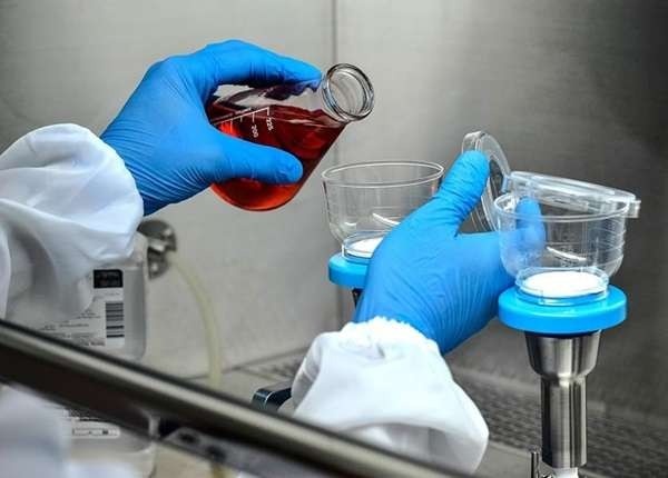
Solid tumors are dissociated using mechanical disruption and enzymatic digestion (e.g., collagenase, DNase) under controlled conditions to release immune cells. Enrichment techniques often follow dissociation: density gradient centrifugation separates mononuclear cells, while red blood cell lysis removes erythrocytes prior to sorting.
Cell sorting: magnetic and flow cytometry approaches
Magnetic-activated cell sorting (MACS) and fluorescence-activated cell sorting (FACS) are standard for isolating macrophage populations. MACS offers rapid positive or negative selection using antibody-coated beads; FACS allows high-resolution sorting based on multiple markers (for example, CD45+ to gate leukocytes, then CD68, CD163, CD206 or F4/80 in mice). Quality control should include viability staining and transcript or protein checks.
Molecular phenotyping
Phenotyping relies on flow cytometry panels, immunohistochemistry (IHC), multiplex immunofluorescence, bulk and single-cell RNA sequencing, and proteomic assays. Spatial transcriptomics and multiplexed imaging link macrophage states to tissue architecture and local cell interactions.
Functional assays and experimental models
In vitro functional assays
Common assays measure phagocytosis (e.g., uptake of labeled beads or apoptotic cells), cytokine secretion (ELISA, multiplex bead arrays), antigen presentation capacity, and chemotaxis. Co-culture systems with tumor cells, fibroblasts, or endothelial cells assess how macrophages influence proliferation, invasion, and drug sensitivity.
In vivo and ex vivo models
Murine tumor models (syngeneic, genetically engineered, or xenograft) permit study of macrophage recruitment and manipulation using genetic tools or pharmacologic inhibitors. Ex vivo tumor slice cultures and organotypic models retain microenvironmental context and can be used for short-term functional testing.
Translational and clinical considerations
Targeting macrophage recruitment and function is an active area of clinical research. Pathways such as CSF1/CSF1R and CCR2 have been explored in trials to modulate TAM abundance or phenotype. Biomarker-driven stratification using immunohistochemistry or gene-expression signatures supports interpretation of therapeutic outcomes. Regulatory oversight for human tissue research includes institutional review boards and national guidelines (e.g., U.S. NIH and equivalent agencies internationally).
For authoritative background on tumor immunology and immune cell roles, consult the National Cancer Institute for resources and guided summaries: National Cancer Institute.
Best practices and reproducibility
Standardization and reporting
Reproducibility benefits from detailed reporting of tissue handling, digestion protocols, sorting gates, antibody clones, and data-processing steps for sequencing and imaging. Inclusion of technical replicates, appropriate controls, and public sharing of raw data and metadata improves cross-study comparisons.
Limitations and interpretation
Ex vivo manipulations can alter macrophage states; enzymatic digestion may cleave surface markers and sorting can stress cells. Spatial context is frequently lost after dissociation, so integrating in situ approaches (IHC, spatial transcriptomics) provides complementary information.
FAQ
What are macrophages in cancer and why do they matter?
Macrophages in cancer, commonly referred to as tumor-associated macrophages (TAMs), influence tumor progression, immune suppression, and therapy response by secreting cytokines, remodeling extracellular matrix, and interacting with other immune and stromal cells. Their heterogeneity and plasticity make them both biomarkers and potential therapeutic targets.
How are macrophages isolated from tumor tissue?
Isolation typically involves mechanical and enzymatic tissue dissociation, enrichment by density gradients, and selection using MACS or FACS based on surface markers. Protocol details vary by tissue type and downstream assays.
Which assays reveal macrophage function in tumors?
Functional assessment includes phagocytosis assays, cytokine profiling, co-culture assays with cancer cells, migration assays, and in vivo depletion or modulation studies in animal models. Single-cell and spatial methods reveal functional heterogeneity within the tumor microenvironment.
Can macrophage states be changed therapeutically?
Preclinical models and clinical trials have explored agents that inhibit macrophage recruitment (e.g., CCR2 pathway) or alter activation (e.g., CSF1R inhibitors). Outcomes depend on tumor type, timing, and combination with other therapies. Ongoing research aims to refine strategies based on molecular subtypes and biomarkers.
Where can researchers find guidelines for working with human tumor tissue?
Researchers should follow institutional review board (IRB) approvals and national regulations for human subjects research, and adhere to best-practice reporting standards for sample handling, data annotation, and sharing. Regulatory agencies and academic consortia provide guidance on ethical and technical aspects of tissue research.