Crop Disease Identifier Guide: Visual Diagnosis Workflow, Checklist, and Best Practices
-

- March 25th, 2026
- 124 views

FREE SEO Topical Map Generator: Find Your Next Content Ideas
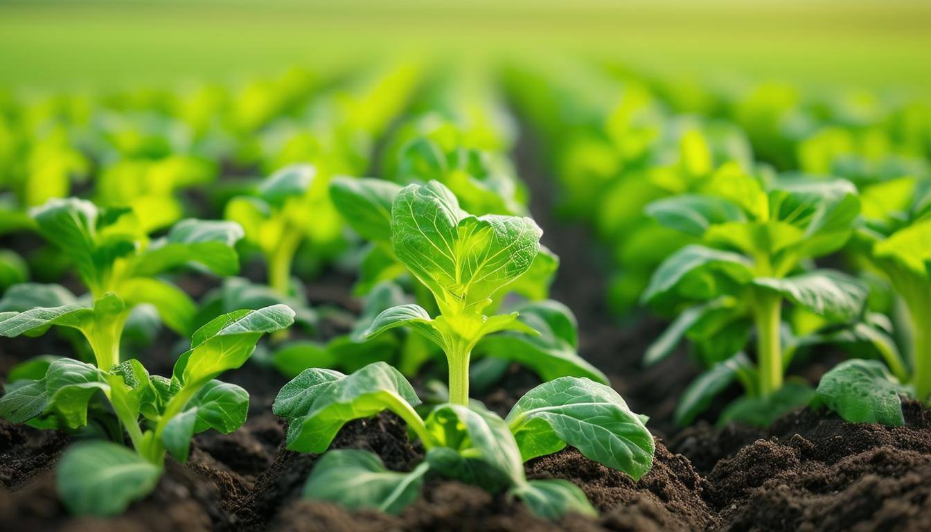
A crop disease identifier turns photos of leaves, stems, fruit, or roots into actionable diagnoses using visual clues and machine analysis. This guide explains how visual disease diagnosis works, which tasks suit automated identification, and practical steps to get dependable results with a plant disease visual diagnosis workflow.
- Understand what a crop disease identifier can and cannot do for visual disease diagnosis.
- Follow the D3 checklist (Detect, Document, Diagnose, Decide, Deploy) for reliable outcomes.
- Use high-quality images, clear metadata, and local verification to reduce misidentification.
How a crop disease identifier works
Image-based systems analyze visible symptoms—chlorosis, necrosis, lesions, pustules, or discoloration—using pattern recognition and trained models such as convolutional neural networks. Many pipelines combine classical image processing (color, texture, shape features) with machine learning classifiers. Some solutions add multispectral or thermal imagery to reveal stress not visible to the naked eye. The process requires labeled datasets, a validation set with confirmed diagnoses, and attention to cultivar and environment variation.
When to use visual diagnosis and when to escalate
Visual diagnosis is effective for common foliar diseases, early blight, rusts, mildews, and nutrient deficiency patterns that produce diagnostic marks. Escalate to lab testing or plant pathology services when symptoms are unclear, when systemic pathogens (e.g., viruses or soil-borne fungi) are suspected, or when biosecurity risks exist. Regulatory guidance from organizations like the FAO is relevant for quarantine and pest management decisions: FAO guidance on pest and disease management.
D3 Checklist: practical framework for visual disease diagnosis
Use the D3 Checklist to standardize image capture and decision-making:
- Detect — Note suspect plants and visible symptoms early.
- Document — Capture multiple images (close-up of lesions, whole plant, affected canopy) with metadata: date, GPS, variety, growth stage.
- Diagnose — Run images through the crop disease identifier, then cross-check outputs against symptom guides and local extension resources.
- Decide — Choose management: monitor, apply targeted control, or submit samples for lab confirmation.
- Deploy — Implement control measures and record results for follow-up monitoring.
Practical tips for reliable plant disease visual diagnosis
- Provide clear, well-lit images: diffuse daylight, avoid heavy shadows, include scale (ruler or coin) and a whole-plant shot plus close-ups of symptoms.
- Capture multiple angles and both healthy and affected tissue to help distinguish disease from abiotic stress.
- Attach context: crop variety, planting date, recent weather, and treatments applied—metadata greatly improves interpretation.
- Use local extension or certified plant clinics to validate any high-risk identification before making regulatory or wholesale treatment decisions.
- Track outcomes: update records when lab confirmation or treatment results are available to improve future accuracy.
Common mistakes and trade-offs
Trade-offs
Speed vs. certainty: automated tools give quick suggestions but can miss rare or symptomatically similar pathogens. Remote sensing covers large areas fast but may miss early, low-coverage infections. Models trained on global datasets may not reflect local cultivar symptoms, creating bias.
Common mistakes
- Submitting low-resolution or out-of-focus photos—models rely on visible detail.
- Relying on a single image or single-shot classification without metadata or context.
- Assuming a confident model output equals definitive diagnosis—confirmation is needed for high-impact decisions.
Short real-world example
A regional agronomist documents yellow-brown lesions on potato leaves during a cool, wet week. Using the D3 Checklist, images are captured (whole plant, lesion close-up) with GPS and weather notes. An image analysis pipeline suggests late blight. The agronomist sends leaf samples to a local plant clinic for lab confirmation. After positive lab results, targeted fungicide application and removal of infected debris reduce spread. Recording the event and photos improves later model retraining for that region.
Implementing or evaluating a leaf disease detection app
Evaluate apps and tools by dataset transparency, validation methods, and ability to export images and metadata for external review. Check whether the provider documents false positive/false negative rates for key pathogens. Prefer solutions supporting local retraining or integration with extension services.
Related terms and concepts
Phytopathology, foliar symptoms, lesion morphology, chlorosis, necrosis, hyperspectral imaging, remote sensing, convolutional neural network (CNN), dataset annotation, plant pathology extension service, integrated pest management (IPM).
Practical next steps
- Start with the D3 Checklist for all suspect cases.
- Collect consistent photos and metadata before running automated tools.
- Validate high-risk or uncertain results with laboratory testing or local experts.
What is a crop disease identifier and how accurate is it?
An image-based tool that compares visual symptoms to known patterns. Accuracy varies by crop, disease, image quality, and training data; reported performance ranges widely and should be interpreted against local validation data.
How should images be captured for the best plant disease visual diagnosis?
Use diffuse natural light, include whole-plant and close-up shots, add scale and context, and record metadata (location, date, variety). Avoid harsh shadows and backlighting.
Can a leaf disease detection app replace laboratory tests?
No. Apps provide rapid preliminary identification and prioritization, but laboratory tests or certified plant health services are required for definitive diagnosis when management or regulatory decisions depend on accuracy.
What is the minimum dataset for reliable disease identification using images?
A reliable dataset should include thousands of labeled images per diagnosis class, representative samples across cultivars and environments, a held-out validation set, and confirmed lab-verified labels when possible.
How to integrate a crop disease identifier into farm operations?
Standardize image capture, route flagged cases to agronomists or extension services, keep records for follow-up, and use confirmed cases to retrain and improve local model performance over time.