Understanding Medullary Sclerosis of the Navicular Bone in Horses
-

- May 20th, 2026
- 3,069 views

FREE SEO Topical Map Generator: Find Your Next Content Ideas
Medullary sclerosis of the navicular bone is a radiographic finding frequently encountered in equine lameness investigations. Although commonly reported in diagnostic imaging, its clinical significance is not always straightforward. In many cases, it may represent either an adaptive response to mechanical stress or an early indicator of more complex podotrochlear disease. Understanding its relevance requires careful interpretation alongside clinical findings, biomechanics, and additional diagnostic tools.
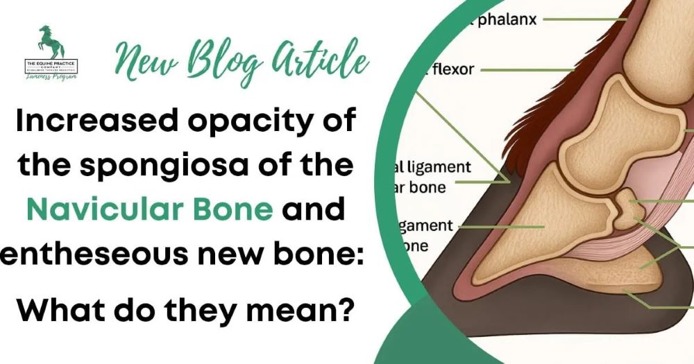
The navicular bone, or distal sesamoid bone, plays a crucial role in equine locomotion. Positioned within the hoof capsule behind the coffin joint, it acts as a fulcrum for the deep digital flexor tendon and contributes to shock absorption during weight-bearing and movement. Because of its anatomical location, it is subjected to high compressive and tensile forces, particularly in performance horses engaged in jumping, racing, or disciplines requiring rapid acceleration and deceleration.
Medullary sclerosis refers to an increase in bone density within the trabecular (spongy) portion of the navicular bone. On radiographs, this appears as increased opacity within the medullary region. This change is believed to result from bone remodeling in response to chronic loading or altered biomechanics within the hoof.
What Causes Medullary Sclerosis in the Navicular Bone?
The development of medullary sclerosis is multifactorial and is primarily associated with chronic mechanical stress. Repetitive loading of the navicular apparatus can stimulate bone remodeling, resulting in increased density over time. This process is similar to adaptive changes seen in other weight-bearing bones of athletic horses.
Several contributing factors may play a role:
- Abnormal hoof conformation or imbalance
- Altered biomechanics due to improper trimming or shoeing
- Increased athletic workload or repetitive strain
- Age-related degenerative changes
- Concurrent soft tissue injury within the podotrochlear apparatus
It is important to recognize that medullary sclerosis does not automatically indicate disease. In some horses, it may represent a normal physiological adaptation to increased workload. In others, however, it may be part of a broader degenerative process involving the navicular bone and surrounding structures.
Clinical correlation is essential. Some horses with significant radiographic sclerosis remain sound and perform at high levels, while others with mild changes may show clear signs of lameness. This variability highlights the importance of interpreting imaging findings within the full clinical context.
Clinical Significance and Diagnostic Approach
The clinical importance of medullary sclerosis depends largely on whether it is associated with lameness or other structural abnormalities. Horses with navicular-related pain often present with subtle and progressive signs that can be easily overlooked in early stages.
Common clinical signs include:
- Intermittent or mild forelimb lameness
- Shortened stride length
- Toe-first landing pattern
- Reduced performance or reluctance to work
- Bilateral forelimb involvement in some cases
A complete lameness examination is essential for accurate diagnosis. This typically involves gait evaluation, flexion tests, hoof testing, and diagnostic nerve blocks. Palmar digital nerve blocks are commonly used to localize pain to the foot region, although they do not specifically isolate the navicular bone.
Radiographic evaluation remains a key diagnostic tool. Standard views include lateromedial, dorsopalmar, and skyline projections. The skyline view is particularly useful for assessing the flexor cortex and internal architecture of the navicular bone, where medullary sclerosis is most visible.
However, radiographs have limitations. They primarily assess bone structure and provide little information about soft tissues. In many cases, navicular-related lameness involves not only the bone but also the deep digital flexor tendon, navicular bursa, and supporting ligaments.
Advanced imaging such as magnetic resonance imaging (MRI) has significantly improved diagnostic accuracy. MRI allows for detailed assessment of both osseous and soft tissue structures within the hoof, enabling a more complete understanding of podotrochlear syndrome.
Management and Clinical Considerations
Management of horses with medullary sclerosis depends on clinical presentation rather than radiographic findings alone. Horses that are clinically sound typically do not require treatment but may benefit from preventive strategies aimed at reducing excessive stress on the navicular region.
These may include:
- Corrective trimming and balanced shoeing
- Optimization of hoof-pastern axis
- Controlled exercise programs
- Regular monitoring in athletic horses
In horses showing clinical signs of lameness, treatment is directed toward reducing pain and improving hoof biomechanics. Farriery plays a central role, with the goal of improving heel support, optimizing breakover, and reducing strain on the deep digital flexor tendon and navicular apparatus.
Medical management may include anti-inflammatory medications and, in selected cases, regenerative therapies. Rehabilitation programs focusing on controlled exercise and gradual return to work are often beneficial.
Early identification of navicular changes is critical, as chronic stress can lead to progressive degeneration over time. Regular monitoring and early intervention can help preserve long-term soundness and performance.
Conclusion
Medullary sclerosis of the navicular bone is a radiographic finding that should always be interpreted in context. While it may represent a benign adaptive change in some horses, it can also be associated with early or progressive podotrochlear disease in others. A comprehensive diagnostic approach combining clinical evaluation, imaging, and biomechanical assessment is essential for accurate interpretation and effective management.
👉 Read the full article here:
https://www.theequinepracticecompany.com/medullary-sclerosis-of-the-navicular-bone/